Biomechanics/Neuromuscular
(13) EFFECTS OF A 12-WEEK HIGH-INTENSITY INTERVAL TRAINING ON VASTUS LATERALIS AND RECTUS FEMORIS CROSS-SECTIONAL AREA AND ECHO INTENSITY IN OLDER ADULTS


April E. Krywe (she/her/hers)
Undergraduate
Creighton University
Omaha, Nebraska, United States
Blake R. Murphy
Research Technician
Creighton Unversity
Omaha, Nebraska, United States
Devon Stoffel (she/her/hers)
Student
Creighton University
Omaha, Nebraska, United States- KH
Kelley Hammond
Assistant Professor
Creighton University
Omaha, Nebraska, United States - JS
Jacob Siedlik
Associate Professor
Creighton University
Omaha, Nebraska, United States - RH
Rashelle Hoffman
Assistant Professor
Creighton University
Omaha, Nebraska, United States - JE
Joan Eckerson
Exercise Science Department Chair
Creighton University
Omaha, Nebraska, United States 
Mitchel A. Magrini, PhD
Assistant Professor
Creighton University
Omaha, Nebraska, United States
Poster Presenter(s)
Author(s)
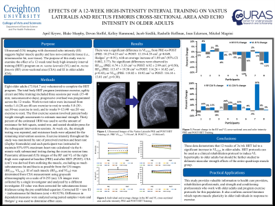
Purpose: Ultrasound (US) imaging with decreased echo intensity (EI) suggests higher muscle quality and less non-contractile tissue (i.e., intramuscular fat, scar tissue). The purpose of this study was to examine the effect of a 12-week total body high-intensity interval training (HIIT) program on m. vastus lateralis (VL) and m. rectus femoris (RF) cross-sectional area (CSA) and EI in older adults (OA).
Methods: Eight older adults (73.8±4.7 yrs) volunteered to complete the HIIT program. The total body HIIT program (resistance exercise, agility circuit, and bike training) included three sessions per week (35-40 min; nonconsecutive days); progressive overload was programmed across the 12-weeks. Work-to-rest ratios were increased from weeks 1-4 (20-sec:40-sec exercise to rest) to weeks 5-8 (30 -sec:30-sec exercise to rest) and to weeks 9-12 (40 -sec:20-sec exercise to rest). The first exercise session involved percent body weight strength assessments to estimate maximal strength. Thirty percent of the estimated 1RM was used to set the amount of resistance for belt squats, seated row, and seated shoulder press for the subsequent intervention sessions. At week six, the strength testing was repeated, and resistance loads were adjusted for the remaining intervention sessions. Exercise intensity throughout the study was monitored by rate of perceived exertion and heart rate (Zephyr biomodule) and each participant was instructed to maintain 85%-95% maximum heart rate calculated via the 6-minute walk submaximal testing during the exercise session time. Panoramic ultrasound (US) images of the RF and VL of the right thigh were captured at baseline (PRE) and after HIIT (POST). CSA (cm2) was derived from outlining the muscle, excluding as much subcutaneous fat and fascia as possible from the US images (RFCSA, VLCSA). EI of each muscle (RFEI, and VLEI) was determined from CSA measurement using grayscale ultrasonography on a scale of 0-255 (au). US images were collected by a single investigator and analyzed by a different investigator. EI value was then corrected for subcutaneous tissue thickness using the pre-established equation: Corrected EI = raw EI + (subcutaneous fat thickness (cm) × 40.5278). Differences in dependent measures were analyzed using paired samples t-tests and Hedges’ g was used to determine effect sizes.
Results: There was a significant difference in VLCSA from PRE-to-POST (PRE: 10.27±4.23 cm2 vs POST: 12.15±4.35 cm2, p=0.049, Hedges’ g=-0.91), with an average increase of 1.89 cm2 (95% CI: 0.002, 3.77). No significant differences were observed in RFCSA (PRE: 6.74 ± 3.33 cm2 vs POST: 6.92 ± 2.99 cm2, p=0.50), RFEI (PRE: 115.47 ± 19.58 cm2 vs POST: 114.26 ± 16.82 cm2, p=0.43), or VLEI (PRE: 118.82 ± 10.92 cm2 vs POST: 116.14 ± 13.01 cm2, p=0.30).
Conclusions: These data demonstrate that 12-weeks of 3x/wk HIIT led to a significant increase in VLCSA in older adults. HIIT protocols can be used as a clinical rehabilitation protocol to induce VL hypertrophy in older adults but should be further studied to delineate muscular strength effects of the entire quadriceps muscle group. Practical Applications: This study provides valuable information to health care providers, rehabilitation professionals, and strength and conditioning professionals who work with older adults and program exercise protocols for this population. It also confirms current literature which reports muscle plasticity in older individuals in response to exercise.
Acknowledgements: None
