Biomechanics/Neuromuscular
(4) TEST-RETEST RELIABILITY OF A 3-D PRINTED ULTRASOUND GUIDE


Carina M. Velasquez, B.S. Ed
Graduate Student
Texas Tech University
Lubbock, Texas, United States
Kealey J. Wohlgemuth, MA, CSCS,*D, CISSN
Graduate Part-Time Instructor
Texas Tech University
Lubbock, Texas, United States- JM
Jacob A. Mota
Assistant Professor
Texas Tech University
Lubbock, Texas, United States
Poster Presenter(s)
Author(s)
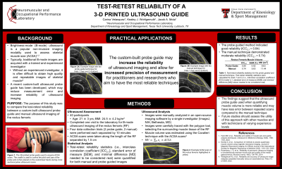
Brightness-mode (B-mode) ultrasound is a popular, non-invasive imaging modality used to assess skeletal muscle size. While popular, it is paramount that these images are acquired with a trained and experienced investigator. Otherwise, it is difficult obtain high quality and repeatable images of skeletal muscle. A recent custom-built ultrasound probe guide has been developed, which may reduce measurement error and increase reliability of ultrasound imaging.
Purpose: The purpose of this study was to compare the test-retest reliability between a custom-built ultrasound probe guide and manual ultrasound imaging for the rectus femoris.
Methods: 40 participants (mean ± SD: age = 21 ± 3 yrs; BMI = 24.5 ± 4.2 kg/m2) completed one visit to the laboratory where each participant underwent non-invasive ultrasound imaging on their rectus femoris (RF) across four separate image acquisition sessions, each separated by 10 minutes. Two trials utilized the custom-built, 3-D printed probe guide while the other two trials did not use a probe guide. The probe guide consisted of a customized cradle which was printed to fit the exact dimensions of the ultrasound probe used in this study. To acquire images, the cradle, which controlled the pitch and yaw of the probe, was then placed on custom-designed tracks while it glided on tracks during a cross-sectional plane ultrasound scan. Anatomical cross-sectional area (ACSA) was quantified every 1.5 cm along the length of the rectus femoris, which was standardized based off of the capabilities of the custom-built probe guide. During the two trials which did not employ a probe guide, scans occurred in the same locations as the aforementioned trials. ACSA was quantified by an experienced investigator carefully selecting the muscle fascia of the rectus femoris using open-source software. Muscle volume was estimated using the Cavalieri technique with the ACSA scans. Test-retest reliability statistics (i.e., interclass correlation coefficients [ICC] model 2,1, standard error of measure [SEM], and the minimal difference [MD] values needed to be considered real) were quantified.
Results: The probe guided method indicated good reliability (ICC2,1 = 0.84, SEM = 4.92, MD = 9.63 cm3) while the ultrasound technique without a probe guide demonstrated moderate reliability (ICC2,1 = 0.74, SEM = 5.83, MD = 11.44 cm3).
Conclusion: The findings from the present investigation suggest that the ultrasound probe guide used when quantifying muscle volume is more reliable and may have less error between repeated scans compared to not having a probe guide. Future studies should assess the utility of this approach with other muscles and with technicians of varying experience levels.
PRACTICAL APPLICATIONS: The custom-built ultrasound probe guide may increase the reliability of ultrasound imaging and may allow for increased precision of measurement for practitioners or researchers who aim to have the most reliable imaging practices.
Acknowledgements: None
